


Video: Horses neck (cervical spine) perfectly x-rayed
plus VAT
This is what awaits you
The e-learning video “Horsecervical spine perfectly X-rayed” is part of our video series “Horses perfectly X-rayed”
Aim of the e-learning video series:
The three-part e-learning video series "Perfectly X-Rayed Horses" offers comprehensive guidance to enhance your equine X-ray diagnostic skills. You'll receive detailed information on precise patient positioning, optimal alignment of the X-ray tube and detector, and an in-depth presentation of modern imaging techniques.
The video series demonstrates various X-ray projections, allowing you to gain precise insights into equine X-ray practice.
Further details can be found here .
Topics of the 3-part video series:
- Horseback perfectly x-rayed
- Horse's head perfectly x-rayed
- Cervical spine perfectly x-rayed
Aim of the e-learning video: “Perfect X-ray of the cervical spine”:
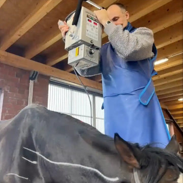
Learn everything you need to know about outpatient X-ray diagnostics of the cervical spine in our e-learning video. Learn all about the necessary tools for standardized preparation, patient positioning, and precise alignment of the X-ray tube and X-ray detector.
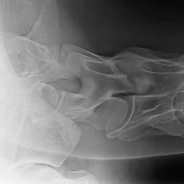
Numerous sample images illustrate the necessary projections and show how they can be created precisely. Typical pathologies are also demonstrated and discussed in detail. This will help you use your X-ray machine specifically and effectively!
Content
E-Learning Video: Cervical spine X-ray
Topics “Perfect X-ray of the horse’s cervical spine”:
General requirements for cervical spine radiography
o Standardized preparation and positioning of the patient
o Technical aids for the exact positioning of X-ray tube and X-ray detector
Standard images for general radiological assessment and topographical representation of the cervical spine
Special radiological recording of the following regions of interest:
o Atlanto-occipital joint
o Articular processes and their joints (facet joints), C2/C3 to C7/T1
o Intervertebral foramina
o Ventral and dorsal tubercles of the transverse processes; ventral plate of C6
o Ventro-lateral surfaces and edges of the vertebral bodies and cranial / caudal extremities.
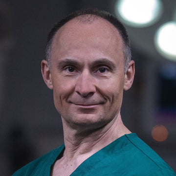
Your speaker
FTA for surgery / FTA for horses / authorization for further training for both specialist areas
Dr. med. vet. Aleksandar Vidovic
Short CV
August 2021 Founding of “Vidocq Equine Surgery Consulting GmbH”
May 2013 - August 2021 Managing Director and Medical Director / from December 2019 Medical Director of the St. Georg Horse Clinic in Trier
August 2011 to May 2013 Lead surgeon at the Burg Müggenhausen horse clinic
Since October 2002 authorization to provide further training in the fields of “surgery” and “horses”
August 2000 Recognition as a specialist veterinarian for horses
March 1999 Recognition as a specialist veterinary surgeon in surgery
1998-2011 Worked in a veterinary clinic for horses in Altforweiler
1995-1998 Assistant doctor
1997 dissertation
1992-1995 surgical training at the Hochmoor Animal Clinic
1985-1991 Studied veterinary medicine at the Faculty of Veterinary Medicine at the University of Belgrade
That’s why you should watch the e-learning video series “Horses perfectly x-rayed” from OstseeCollege
Valuable knowledge offering with many advantages:
The OstseeCollege’s e-learning video series offers in-depth insights into the world of perfect X-rays – a valuable source of knowledge with many advantages:
Flexibility: 24/7 access for flexible learning according to your schedule
Repetitions: Unlimited repetition options to deepen understanding
Self-paced: Learn at your own pace without time constraints
Location independence: Access from anywhere for a flexible learning environment
Long-term access: Permanent access allows you to refresh or deepen your knowledge over the long term
X-raying horses with a practice-oriented approach
Our video series provides you with the latest scientific findings in imaging techniques that you can apply in your daily practice. In this three-part video series, we cover the following topics:
- X-ray of the cervical spine in horses
- Horseback X-rays: mobile / outpatient and inpatient
- Horse head x-ray
X-raying horses: From experts for experts
Our speakers are experienced veterinarians specializing in surgery and equine veterinary physiotherapy. Their many years of experience enable them to bring a wealth of practical examples to their veterinary X-ray courses, which is why we recommend the complete video series on horse X-rays.


